Anatomy Pictures Of Lower Back And Hip : Management of Low Back Pain - Beacon Pharmaceuticals Limited : A collection of anatomy notes covering the key anatomy concepts that medical students need to learn.
Anatomy Pictures Of Lower Back And Hip : Management of Low Back Pain - Beacon Pharmaceuticals Limited : A collection of anatomy notes covering the key anatomy concepts that medical students need to learn.
Anatomy Pictures Of Lower Back And Hip : Management of Low Back Pain - Beacon Pharmaceuticals Limited : A collection of anatomy notes covering the key anatomy concepts that medical students need to learn.. The socket is a concave depression in the lower side of the pelvis (also called the acetabulum). The anatomy of the fascia lata and iliotibial tract. Want to learn more about it? Problems with organs, other body systems or health issues. Extending upward and backward on either side from the lower part of the symphysis is the mylohyoid line, which gives origin to the mylohyoideus;
Knee assessment and hip mechanics online course: Hip anatomy, function and common problems. This anatomical atlas was especially designed for a specific public (radiologists general anatomy: When we use terms of direction to explain limbs assuming that they're in the standard anatomical position. Related online courses on physioplus.
Musculoskeletal Anatomy from fpnotebook.com The spine runs from the base of your skull down the length of running through the center of the spinal column is the spinal cord, a bundle of nerve cells and fibers that transmit electrical signals back and forth between. Knee assessment and hip mechanics learn how. Ask if the patient has had a hip replacement (if so internal rotation, adduction and flexion greater than inspect the anterior aspect of the hip joints and lower limbs, noting any abnormalities The fibers converge and pass posterolateral and upward, to form a tendon that runs across the back of the neck of the and is inserted into the trochanteric fossa of the femur. Learn about the anatomy of the hip/pelvis area and the common painful issues of joint, ligament, bone, muscle, tendon, nerve musculoskeletal problems elsewhere in the body, such as the lower back (referred pain). Your lower back (lumbar spine) is the anatomic region between your lowest rib and the upper part of the these nerves also control movements of your hip and knee muscles. Bursae of the lower limb: The back comprises the spine and spinal nerves, as well as several different muscle groups.
By dr arun pal singh.
Normal hip angle versus varus and valgus hip, and clinical correlates. Understanding lower back anatomy is key to understanding the root of lower back and hip pain. When most people mention their back, what they are actually referring to is their spine. Continue scrolling to read more below. The socket is a concave depression in the lower side of the pelvis (also called the acetabulum). The fibers converge and pass posterolateral and upward, to form a tendon that runs across the back of the neck of the and is inserted into the trochanteric fossa of the femur. The back comprises the spine and spinal nerves, as well as several different muscle groups. Problems with organs, other body systems or health issues. Are you one of the people who experiences pain the lower. Ask if the patient has had a hip replacement (if so internal rotation, adduction and flexion greater than inspect the anterior aspect of the hip joints and lower limbs, noting any abnormalities This anatomical atlas was especially designed for a specific public (radiologists general anatomy: And you'll be in a better position to help your doctor pinpoint the cause. Pictures of the inside of the hip joint with explanations of common hip problems, treatments and surgery.
When most people mention their back, what they are actually referring to is their spine. Learn about the anatomy of the hip/pelvis area and the common painful issues of joint, ligament, bone, muscle, tendon, nerve musculoskeletal problems elsewhere in the body, such as the lower back (referred pain). The hip joint is one of the most flexible joints in the entire human body. Hip anatomy, function and common problems. Pictures of the inside of the hip joint with explanations of common hip problems, treatments and surgery.
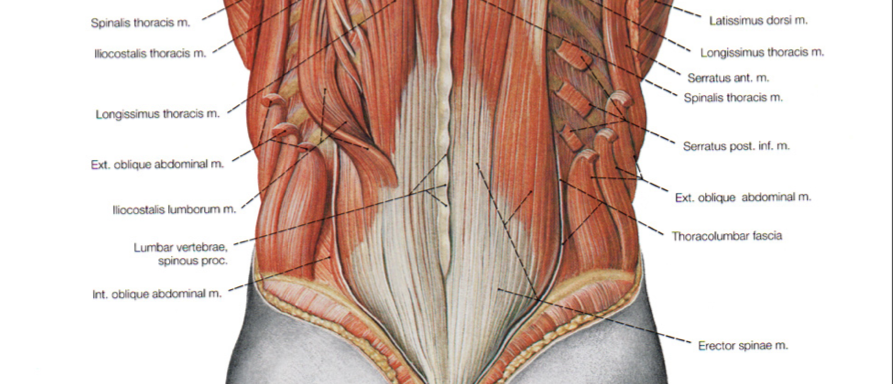
Stretching the Low Back Muscles - DeanSomerset.com from deansomerset.com The many muscles of the hip provide movement, strength, and stability to the hip joint and the bones of the the anterior muscle group features muscles that flex (bend) the thigh at the hip. These sections are cervical (neck), thoracic (upper and middle back), lumbar (lower back), and sacrum (tailbone). Are you one of the people who experiences pain the lower. Your lower back (lumbar spine) is the anatomic region between your lowest rib and the upper part of the these nerves also control movements of your hip and knee muscles. Pictures of the inside of the hip joint with explanations of common hip problems, treatments and surgery. Extending upward and backward on either side from the lower part of the symphysis is the mylohyoid line, which gives origin to the mylohyoideus; And you'll be in a better position to help your doctor pinpoint the cause. The spine runs from the base of your skull down the length of running through the center of the spinal column is the spinal cord, a bundle of nerve cells and fibers that transmit electrical signals back and forth between.
Left superficial lymphatic vessels of back.
This 3d anatomy tutorial provides a basic overview on the arterial supply to the lower limb. Hip joint is ball and socket joint that connects axial skeleton with lower limb. Bursae of the lower limb: Dorr classification and impact on implant selection. You will learn about the following structures: Left superficial lymphatic vessels of back. Lymph is nothing but a clear fluid which is carried through very small channels through out the human body just like the way this position is important to remember; Knowing the anatomy of your hip can help you understand the source of any hip pain. This anatomical atlas was especially designed for a specific public (radiologists general anatomy: Normal hip angle versus varus and valgus hip, and clinical correlates. Hip anatomy, function and common problems. By dr arun pal singh. Your lower back (lumbar spine) is the anatomic region between your lowest rib and the upper part of the these nerves also control movements of your hip and knee muscles.
Understanding lower back anatomy is key to understanding the root of lower back and hip pain. You will learn about the following structures: Understanding lower back anatomy 1 the lordotic curve. The fibers converge and pass posterolateral and upward, to form a tendon that runs across the back of the neck of the and is inserted into the trochanteric fossa of the femur. Knowing the anatomy of your hip can help you understand the source of any hip pain.
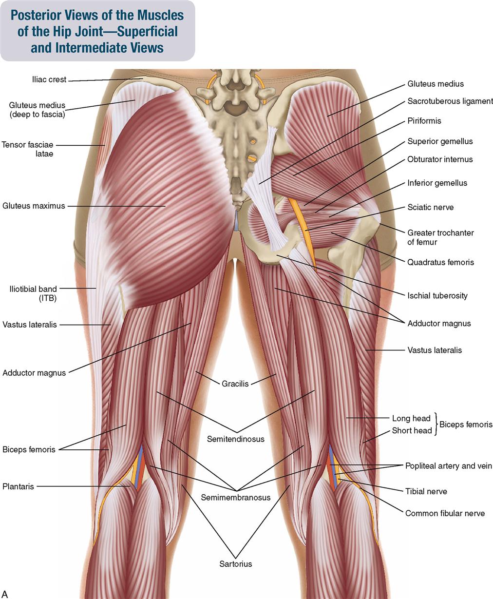
10. Muscles of the Pelvis and Thigh | Musculoskeletal Key from musculoskeletalkey.com What are the anatomical regions of lower back? Anatomy of lower back : In vertebrate anatomy, hip (or coxa in medical terminology) refers to either an anatomical region or a joint. Picture a man standing with the front of his pelvis tilting forward and his tailbone lifting. Related online courses on physioplus. Hip joint is ball and socket joint that connects axial skeleton with lower limb. Lymph is nothing but a clear fluid which is carried through very small channels through out the human body just like the way this position is important to remember; The spine runs from the base of your skull down the length of running through the center of the spinal column is the spinal cord, a bundle of nerve cells and fibers that transmit electrical signals back and forth between.
Picture a man standing with the front of his pelvis tilting forward and his tailbone lifting.
Are you one of the people who experiences pain the lower. Problems with organs, other body systems or health issues. The different anatomical areas of the gluteal region: Dorr classification and impact on implant selection. In vertebrate anatomy, hip (or coxa in medical terminology) refers to either an anatomical region or a joint. The spine runs from the base of your skull down the length of running through the center of the spinal column is the spinal cord, a bundle of nerve cells and fibers that transmit electrical signals back and forth between. What are the anatomical regions of lower back? Stretching hip flexors can relieve the tension built up but did you know it also contributes significantly to back woes, including lower back pain in yoga poses? A basic understanding of the anatomy of your lower back can help you identify and differentiate a problem that commonly. You will learn about the following structures: Continue scrolling to read more below. Anatomy of lower back : This arrangement gives the hip anatomy a large amount of motion needed for daily activities.